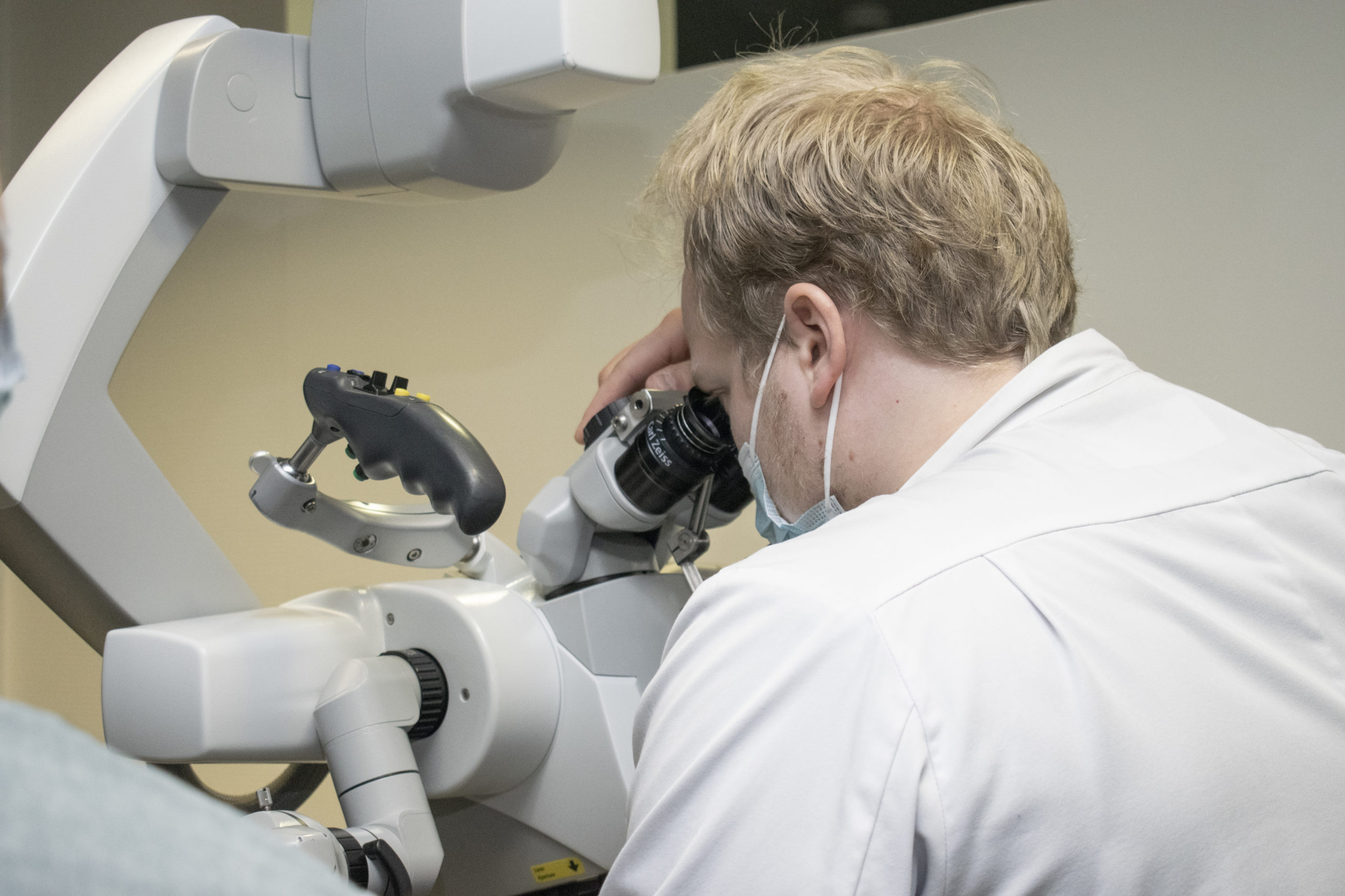

Doctoral researcher Sami Puustinen is part of a study aimed at analysing the potential of using hyperspectral imaging to, in particular, distinguish between functionally important tissue types. It can be difficult to tell nerves apart visually during cancer surgery, and removing nerve tissue with the cancerous tissue can leave the patient with permanent paralysis.
The study relies on a hyperspectral camera developed by Senop and involves teams of researchers from the University of Eastern Finland designing software to process data relating to the optical properties of different tissue types. Professor of Neurosurgery Juha E Jääskeläinen from the University of Eastern Finland hopes to incorporate spectral imaging technology into all microscopes and endoscopes used in surgery as soon as possible.
“Senop plays a major role in the study. This partnership is critical to our ability to disseminate the new technology among neurosurgery specialists.”
Sami Puustinen, Researcher
“The more data about living tissue we input into the system, the faster the system learns. Our plan is to compile a surgical spectral imaging databank to ultimately share with others as well”, he says.
“Senop plays a major role in the study. This partnership is critical to our ability to disseminate the new technology among neurosurgery specialists. Docent Elomaa’s work in the medical applications of photonics is groundbreaking in Europe, and we are grateful for his contribution”, he adds.