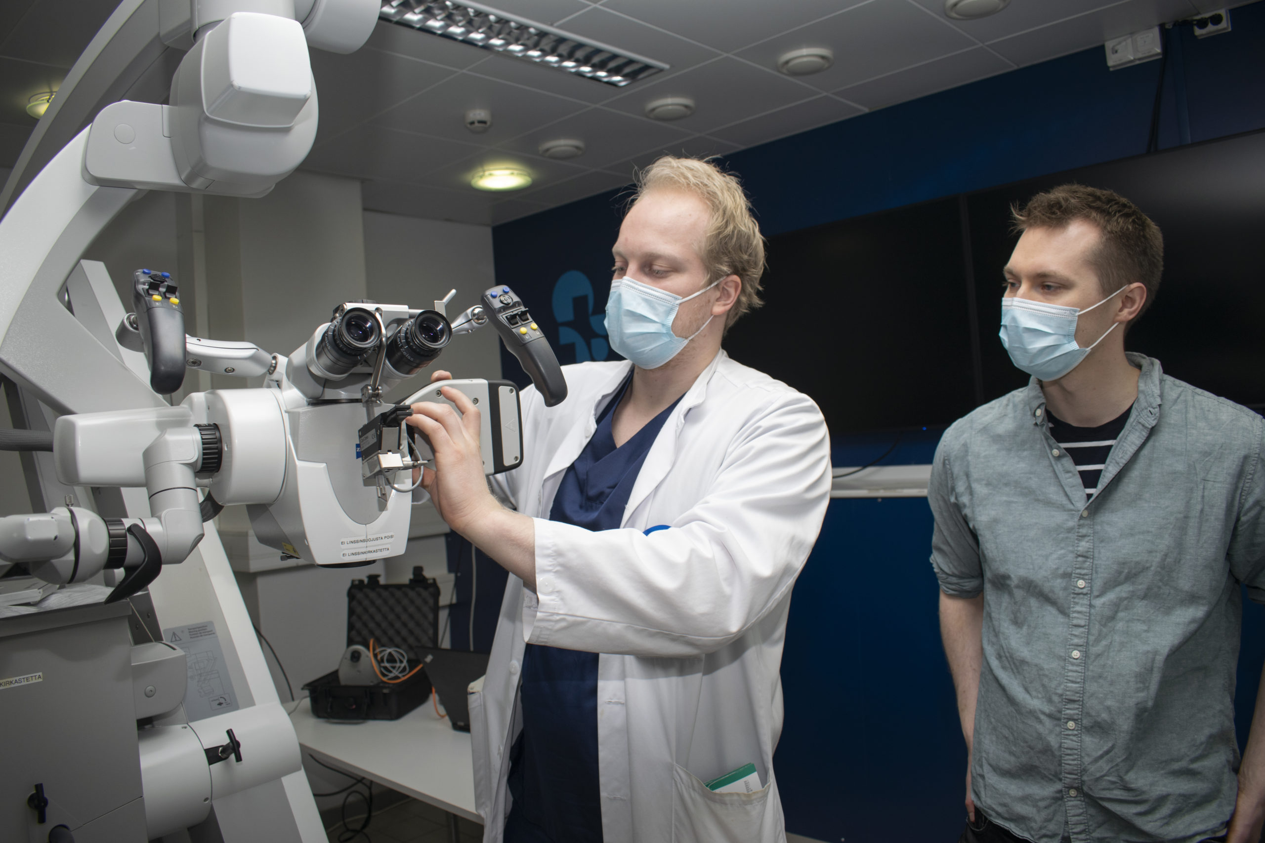

Senop and the Kuopio University Hospital’s Microsurgery Centre have a long history of collaboration in the field of medical research on hyperspectral imaging. By iterating together, i.e. by jointly developing the product in stages, they have come very close to a solution that can be utilised in surgeries. The HSC-2 hyperspectral camera is well suited to imaging organic materials, and it has around one terabyte of memory.
“There is really a lot of potential for utilisation in medicine, whether at issue is the imaging of cancer tumours or functionally important tissues that are to be spared in the operation.”
Antti-Pekka Elomaa, Docent of Neurosurgery
An integral part of future operating room equipment
The Microsurgery Centre is a meeting place for clinical medicine professionals, researchers and technology companies. Spectral imaging, 3D modelling and eye-tracking technologies improve the surgeon’s ability to visualise brain and tissue structures and plan the procedures to be performed. There are both surgical operating microscopes and endoscopic instruments available that are used in endoscopic surgery.
In 2017, Senop’s solution won the competitive tendering for cameras carried out by the Microsurgery Centre as part of a project of the Region of North Savo and the European Regional Development Fund related to hyperspectral cameras. Current research projects, such as those funded by the Finnish Medical Foundation, are examining the pathology of central nervous system tumours in particular.
“The product seemed best suited for clinical use and is a key instrument in the study. Open interfaces are a clear plus.”
“The product seemed best suited for clinical use and is a key instrument in the study. Open interfaces are a clear plus. It is easy to make device customisations together when communication works well”, Elomaa says.
Elomaa believes that, in the future, operating rooms will be equipped with hyperspectral imaging devices, the use of which will improve surgery outcomes regardless of specialty. Spectral imaging is highly complex, so data-driven know-how and innovation play an important role. Senop’s hyperspectral camera is equipped with a C-mount mechanism compatible with several cameras and microscopes.
“There is really a lot of potential for utilisation in medicine, whether at issue is the imaging of cancer tumours or functionally important tissues that are to be spared in the operation.”
Challenges posed by sufficient exposure time
Traditional optical operating microscopes typically have powerful xenon lights, so the exposure time can be set short, and the colour spectrum is rich.
“To an increasing extent, endoscopes may have problems with the sufficiency of light, especially when using narrow-band light sources. An efficient adjustable light source would make an important working pair for a hyperspectral camera”, Elomaa says.
The live functionality that will be incorporated as part of the camera allows the user to select the wavelength ranges to be displayed as well as the exposure time, and the image can be viewed and analysed on the screen in real time.
“It is easy to make device customisations together when communication works well.”
“The live functionality looks really good; it is exactly what we need. Digital enhancement and the ability to expand the image view are important features. Automatic exposure time adjustment would be necessary, as conditions vary during surgery and there is no time to approximate the appropriate exposure time”, Elomaa says.